
0%
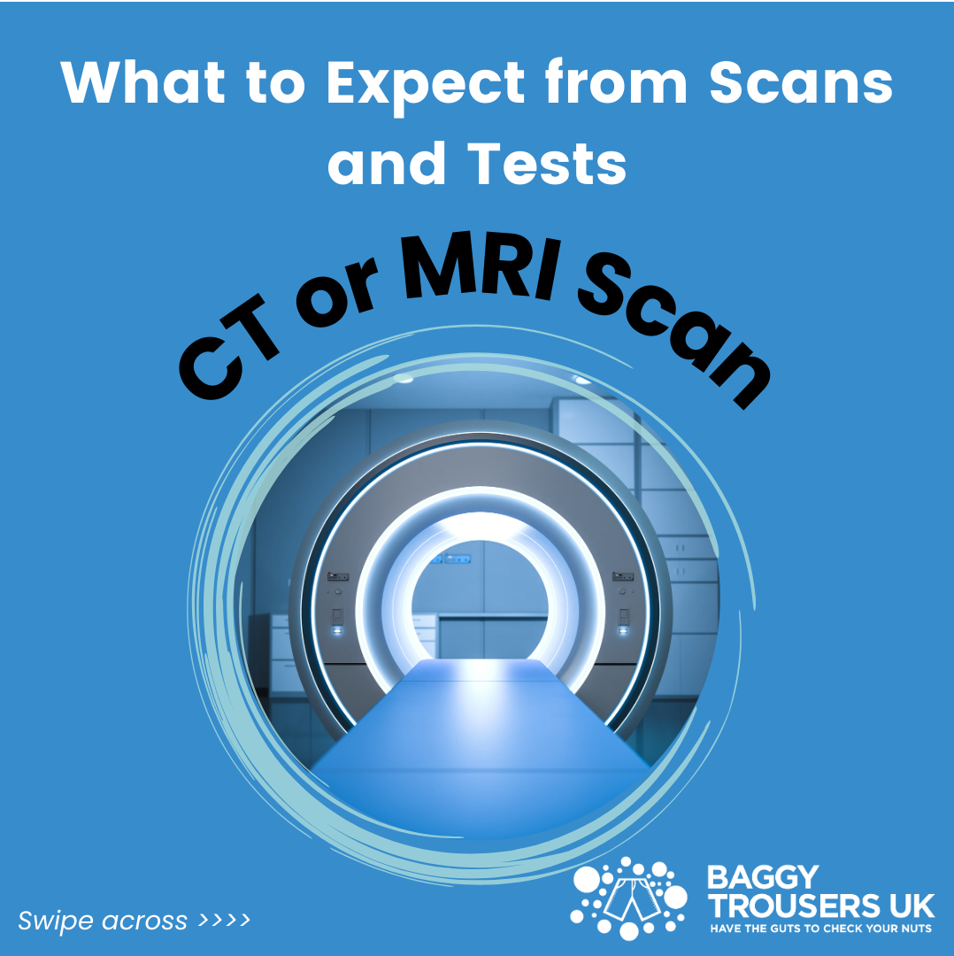
What to Expect from Scans and Tests: CT or MRI Scan
The first few stages of a testicular cancer diagnosis will usually involve an ultrasound scan and blood tests. We covered what you can expect from those tests in the previous blogs in this series. If the combined results of these tests suggest that a lump or abnormality may be cancerous, further investigation will be required.
The next step may involve having a CT (Computed Tomography) or MRI (Magnetic Resonance Imaging) scan to confirm diagnosis, and to check if the cancer has spread. Both of these techniques provide detailed images of the inside of your body, but they work in different ways.
Differences between CT scans and MRI scans
MRI scan: Uses magnetic fields and radio waves to generate images, with no radiation. This method provides highly detailed pictures of soft tissues like the brain, muscles, nerves, and organs.
CT scan: Uses X-rays to create images of the body in cross sections. As the X-ray beams pass through the body, detectors measure the radiation that comes out the other side. This provides clear imagery of bones, organs and blood vessels.
Both types of scan can be used to detect cancer. CT scans are more common for initial diagnosis and staging. They provide a quicker and more comprehensive view of the body, including the detection of tumours and other abnormalities in organs. However, MRI scans are sometimes preferred when dealing with soft tissue cancers, such as testicular cancer.
When do you have a CT or MRI scan?
An ultrasound scan is the first imaging test for detecting testicular cancer. It is the most common and the most effective. If your blood test results show tumour markers, and an ultrasound scan shows abnormalities synonymous with a tumour, the likelihood is that you have testicular cancer. It is at this stage that you will be sent for a CT or MRI scan. The purpose of this is to confirm the diagnosis and to primarily check if the cancer has spread to other parts of your body.
What are the scans looking for?
CT scans are typically used to check for signs that the cancer has spread beyond the testicle. It helps determine the size, stage and location of the tumour, which is crucial for planning your treatment.
A CT scan usually checks your:
- lymph nodes, particularly in the abdomen and pelvis
- lungs or liver
- chest and abdomen
An X-ray may also be used to check your lungs.
MRI scans provide superior imaging of soft tissues, so they are also often used to provide detail on tumours in the testicle, scrotum, or surrounding areas. This helps to investigate cancer that may have spread to nearby tissues, like the spermatic cord or scrotal wall.
Where do you have a CT or MRI scan?
CT and MRI scans are usually conducted at a hospital. However, you may also be referred to a specialist clinic, imaging centre or urgent care centre.
What happens next?
Your test results will be sent to an assigned urologist. They will analyse the results and invite you to an appointment to talk about the next steps. You should hear from them within a matter of days, or no more than a week.
Biopsy is not possible with testicular cancer without first removing the affected testicle in surgery. If all of the test results confirm the signs of cancer, this will be the next step. If your results show cancer has spread, the urologist will also talk to you about the potential treatment plan and what to expect after your surgery.
Read our blog Caregiver Chronicles: Meeting the Urologist for more information on what to expect in your appointment.
Contact us for support
A lot of men find it helpful to talk while going through a testicular cancer diagnoses. This is a really difficult period and one that can impact your mental health massively. Don’t go through it alone. Here at Baggy Trousers UK we have a number of members that have experienced testicular cancer, and they will be more than happy to talk to you.
For more information contact us using our contact form, or get hold of us on Facebook, Instagram, or X (Twitter).